Your Guide to Pterygium Eye Treatment
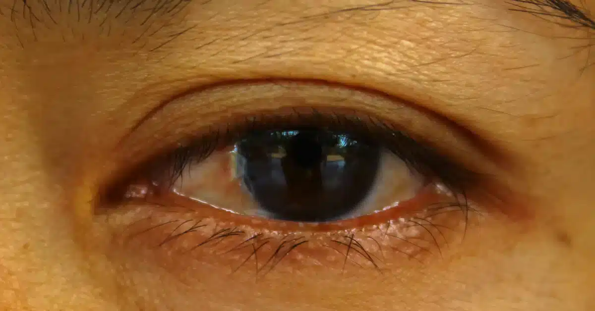
A Pterygium is a fleshy, elevated growth on the eye’s conjunctiva.
The white of your eye is covered by a thin, transparent membrane called the conjunctiva.
Typically, the conjunctiva stops at the cornea, the fine front section of the eye. The interior of your eyelids is lined with it as well.
The term “Pterygium” was coined by combining the Greek words for “wing” (ptéryga) and “fin” (pterýgio).
A Pterygium is a wing- or triangle-shaped growth of extra conjunctival tissue.
In most cases, it develops at the side of the eye closest to the nose but can appear at any corner.
It can even reach the cornea of your eye and spread there.
Although Pterygium can affect either eye, it usually only affects one at a time. But there are two ways in which it can be treated.
It can either be through medications or a surgical process.
Pterygium is considered bilateral when it develops in both eyes simultaneously.
Pterygium Eye Treatment
A Pterygium usually doesn’t need to be treated until it’s obstructing your vision or causing severe discomfort.
You should schedule a regular visit with your eye doctor. This will help him identify if the growth is causing vision problems.
According to the condition, your eye care provider can provide you with specific treatments:
Medications
If the Pterygium is causing you problems like redness or irritation, your eye care doctor may prescribe eye drops or ointments.
These medications contain corticosteroids that help in reducing inflammation.
Surgery
When non-invasive therapies have failed, your doctor may suggest surgical tissue grafting.
Amniotic Membrane Transplantation (AMT) and Conjunctival Autograft (CAG) surgery are two examples of frequently performed surgical procedures.
Conjunctival autografting surgery uses tissue from another eye area and some limbal tissue. It is for covering the place where the Pterygium once was.
Amniotic membrane transplantation has been shown to reduce inflammation and scarring, two of the leading causes of Pterygium recurrence.
The likelihood of a Pterygium returning can be decreased by grafting scleral tissue onto the affected location after surgery.
Another strategy for avoiding a recurrence is using a topical medicine like Mitomycin C (which prevents aberrant tissue growth and scarring).
An eye patch should be worn for the first day or two after a Pterygium excision. Most surgical patients can return to work the day after their procedure.
Patients are advised to use anti-inflammatory steroid eye drops as their eyes heal several months after surgery.
A year following surgery, individuals with Pterygium are constantly examined due to the high risk of recurrence.
Several standard surgical techniques are used for Pterygium removal, but most procedures last about 30 minutes.
Patients should be aware that this surgery sometimes induces or exacerbates Astigmatism.
Diagnosis

Your eye care provider uses a slit lamp to diagnose Pterygium.
A slip lamp is a microscope that focuses a narrow (a “slit”) line of bright light on your eye.
It helps your eye care provider to look at the front and inside of your eye. This whole process is a normal eye examination process.
Other tests performed by your eye care could be:
- Corneal topography: Using this imaging technology, a 3D map of the corneal surface can be generated on a computer.
- Visual acuity test: This test checks your vision clarity from 20 feet apart.
Your eye care specialist can take pictures of your eyes. This can be done to track the growth of Pterygium over time.
Prevention

Pterygium can be avoided by limiting exposure to environmental risk factors such as sunshine, wind, and dust by shielding one’s eyes with UV-blocking sunglasses and a hat.
Using these safeguards following Pterygium removal surgery may reduce the risk of a recurrence of the condition.
Equally effective in preventing Pterygium is eye protection in highly polluted areas.
Conclusion
Although Pterygium can be surgically removed, the effects remain for a more extended period, for which doctors usually prescribe eye drops.
On the other hand, the recurring issue of Pterygium is also treatable by grafting scleral tissue onto the affected location after surgery.
With conjunctival autograft and fibrin glue, Pterygium excision has a low recurrence rate, good cosmetic success, and an acceptable Pterygium surgical time.
Frequently Asked Questions
How do you treat Pterygium in the eye?
Artificial tears and steroid ointment or eye drops could be comforting regarding Pterygium. However, it looks scary and odd, even though the condition is not so severe.
How do you get rid of Pterygium without surgery?
Treatment of Pterygium can be done with the help of steroid eye drops, artificial tears, or ointments with corticosteroids. Your doctor may recommend Pterygium surgery if the growth causes extreme discomfort or begins to impact your vision significantly.
Is Pterygium removal painful?
You can experience discomfort for several days after the surgery. In this case, your doctors may give you eye drops six times a week.
WowRx uses only high-quality sources while writing our articles. Please read our content information policy to know more about how we keep our content reliable and trustworthy.


